
2026年7月深圳湾实验室分子生理学研究所李芬芳课题组及合作者在Journal of Controlled Release 在线发表题为“Cross-scale mechanistic insights into ultrasound pulse-length dependent BBB opening”的研究论文。该研究通过跨尺度BBB模型和成像技术(Figure 1),发现即使在被动空化信号检测(passive cavitation detection (PCD))未侦测到惯性空化的条件下,0.5MPa长脉冲超声仍可通过微泡的“循环微射流”导致脑血管内皮细胞局部脱落,而短脉冲超声则能实现均匀、可逆的血脑屏障开放。同时,研究利用超声造影成像实时监测微泡在脑内的灌注动力学,为超声作用BBB窗口的精准设定提供了直接依据。
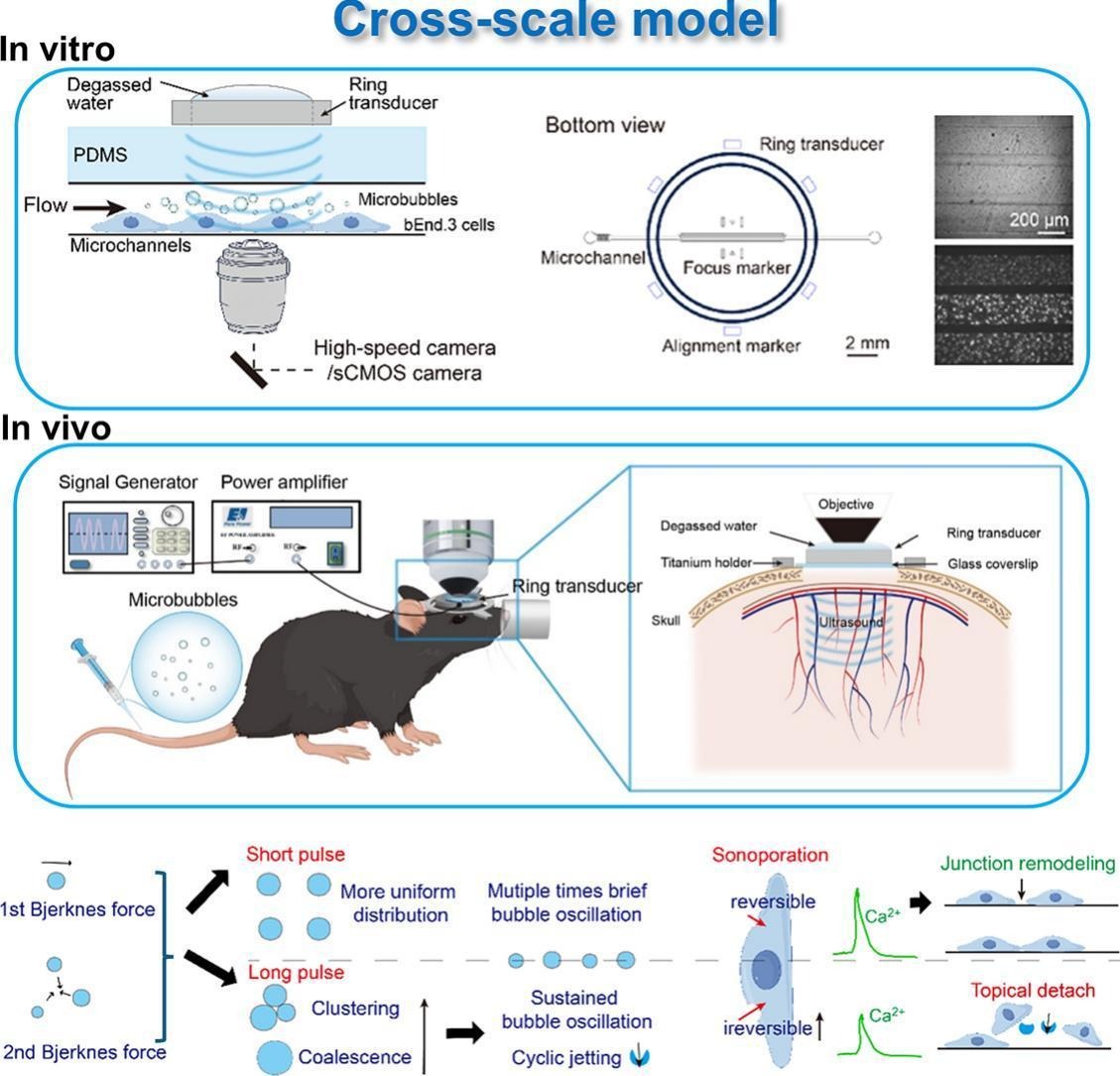
Figure 1. Graphical abstract. The top panels show the experimental methods while the bottom panel summarizes the mechanistic insights gained from this study.
血脑屏障由紧密连接的脑微血管内皮细胞构成,在保护中枢神经系统免受毒素和病原体侵害的同时,也阻碍了绝大多数治疗药物进入脑实质,严重制约了脑肿瘤、阿尔茨海默病、帕金森病等中枢神经系统疾病的药物治疗。聚焦超声联合微泡技术能够无创、可逆地开放血脑屏障,已在全球范围内进入多项临床试验。然而,临床常用的长脉冲方案(毫秒级脉冲)在取得高效递送的同时,也被报道伴有红细胞外渗、短暂性水肿和炎症等副作用。传统观点认为,惯性空化是造成生物损伤的主要元凶,因此科研界普遍通过PCD监测宽带噪声来规避风险。新兴的短脉冲序列虽展现出更宽的安全窗口,其背后的生物物理机制缺乏直接的微观证据。
针对这一科学问题,研究团队建立了一套跨尺度研究体系。在体外层面,利用模拟脑血管的微流控芯片,以25,000帧/秒的高速成像实时捕捉微泡动力学行为,同时通过荧光成像同步监测内皮细胞的声穿孔、钙信号和细胞脱落等生物效应;在活体层面,借助双光子成像技术,在颅窗手术的小鼠脑中实时观察血脑屏障开放的全过程。
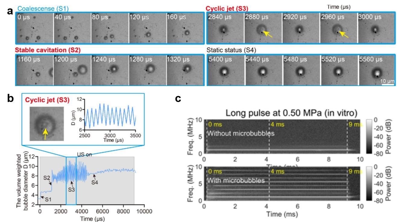
Figure 2. Bubble dynamics inside the microchannel under long pulse 0.5 MPa acoustic pressure. (A) High-speed recordings of individual microbubbles under long-pulse ultrasound exposure (t = 0–9090 μs) at 0.5 MPa acoustic pressure. The bubble kept oscillating from 1240 μs (stable cavitation) when generated from the coalescence of two bubbles till 4400 μs before it merged with another bubble. Cyclic jetting is observed during stable oscillation. (B) Evolution of the diameter of the microbubbles shown in (A) during long pulse ultrasound exposure. (C) Passive Cavitation Detection (PCD) analysis.
研究团队在体外微流控血管模型中实时追踪微泡(初始平均直径2-4 µm)动力学与内皮细胞响应。结果显示长脉冲超声(脉冲长度9.09 ms)驱动微泡在一阶和二阶声辐射力下移动、聚集、融合,形成与换能器频率共振的较大气泡,并在随后的稳定振动中反复产生朝向壁面的“循环微射流” (Figure 2 a-b)。这种循环微射流产生的局部剪切应力比其他机制至少高一个数量级,可造成内皮细胞不可逆声穿孔和局部片状脱落(Figure 3)。值得注意的是,PCD频谱仅显示清晰的谐波峰而无明显宽带噪声,表明体系处于稳态空化主导区 (Figure 2c)。这意味着传统上被认为“安全”的稳态空化,在长脉冲条件下亦可经由循环微射流引发机械损伤。
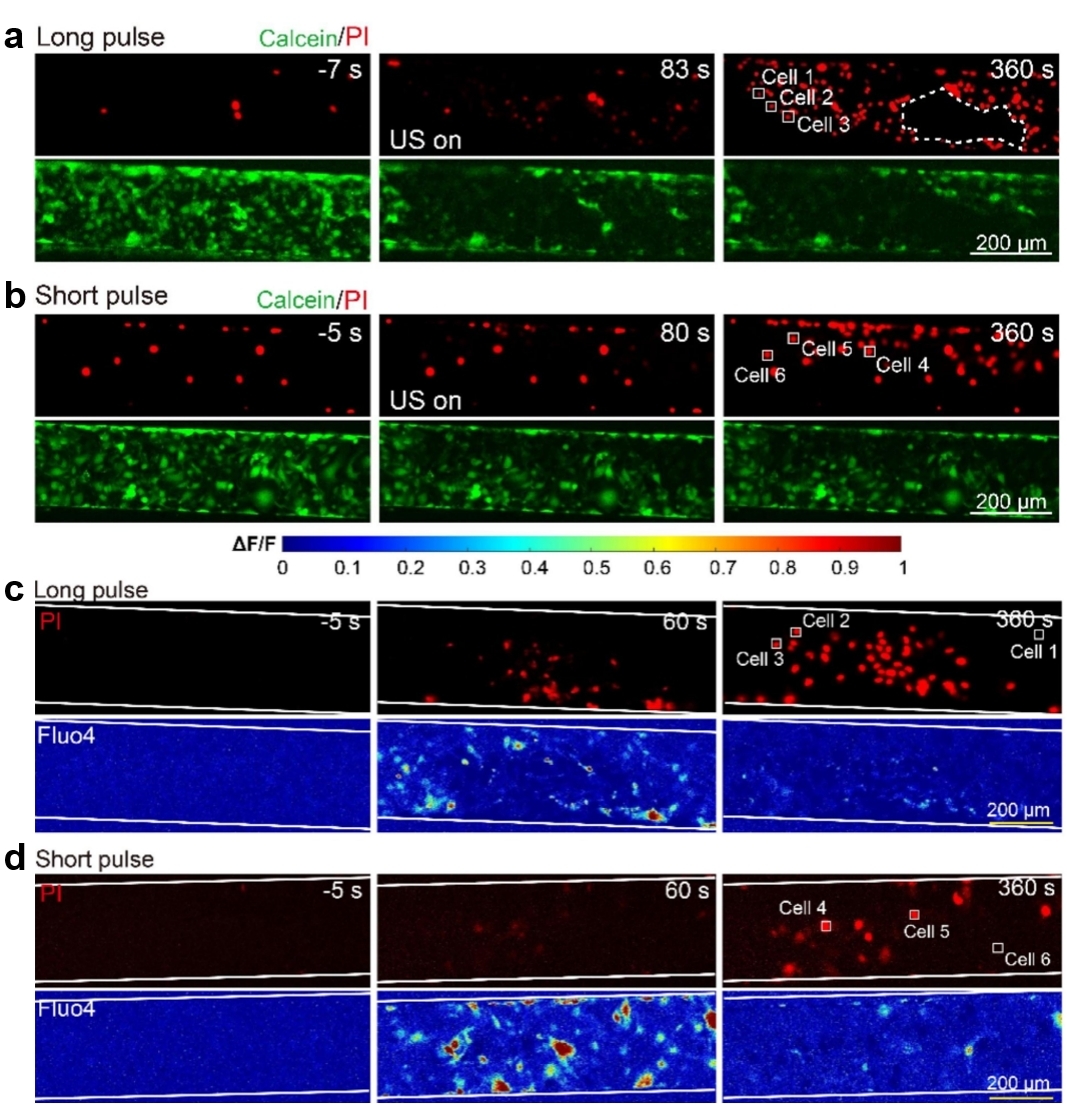
Figure 3. Characteristics of cell monolayer bioeffects under 0.5 MPa ultrasound exposure in the microchannel. (A) and (B) PI uptake (red, upper panel) and calcein leakage (green, bottom panel) in bEnd.3 cell monolayer in the microchannels before (−7 s or − 5 s), soon after (83 s or 80 s), and 360 s after ultrasound exposure at long and short pulse mode, respectively. The dashed line in panel d depicts the outline of the cell detachment region. (C) and (D) PI uptake (red, upper panel) and Ca2+ signaling (pseudo color, bottom panel) in bEnd.3 cell monolayer in the microchannels before (−5 s), during (60 s), and after (360 s) 0.5 MPa ultrasound exposure at long pulse and short pulse mode, respectively. The same pseudo color coding (∆F/F) is used here as in panel c-d.
该研究在体外血管模型中观测到了长脉冲作用下细胞层的局部片状脱落(Figure 3)。细胞脱落可能导致局部血管损伤或破坏,而不均匀、过度的膜穿孔和Ca2+负荷则会导致跨细胞转运不均和细胞凋亡。相反,如果检测不到细胞脱落,同时细胞膜穿孔轻微、分布更均匀,并且Ca2+反应整体性增强,则可通过促进跨细胞转运和在更大血管区域内可逆地广泛开放内皮紧密连接,从而实现更安全的血脑屏障开放。
为验证这一体外发现,团队在活体小鼠脑内进行了双光子实时成像,并结合超声造影成像(Contrast enhanced ultrasound (CEUS))监测微泡的脑灌注时程。 CEUS显示,尾静脉注射的微泡在约60秒达到脑内峰值浓度,半衰期为8-10分钟。该数据指导了后续超声作用时间点和时长(120秒)的设定,确保实验在微泡浓度充足的时间窗口内进行。体内PCD监测和频谱分析仅显示谐波峰而无明显宽带噪声,表明体系同样处于稳态空化主导区 (Figure 4)。
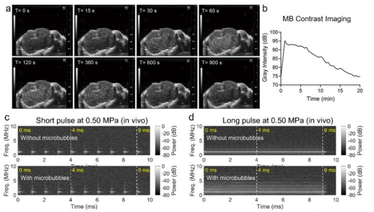
Figure 4. Microbubble contrast imaging and Passive Cavitation Detection measurement at 0.50 MPa pressure in vivo. (A) The ultrasound contrast images of the mouse brain in vivo after a bolus injection of microbubbles through the tail vein. Craniotomy surgery was performed before ultrasound imaging with a 40 MHz transducer. (B) The time-intensity curve of the ultrasound contrast images. Bubbles are injected at t = 0 s. Contrast imaging showing that microbubble concentration in the mouse brain vessels reached peak value at around 60 s and maintained at high level during the sonication period. (C) PCD results for in vivo short-pulse sonication. (D) PCD results for in vivo long-pulse sonication.
在长脉冲作用下,体内双光子成像清晰观察到脑内皮细胞(tdTomato标记)在超声开启后仅9.6秒即发生局部荧光丢失(Figure 5f),且180秒后仍未恢复,直接证实了体内存在内皮细胞脱落。而短脉冲超声(100 μs脉冲,1 kHz重复频率)虽同样能打开血脑屏障,却未造成内皮损伤,且右旋糖酐外渗更为均匀,屏障在数分钟内迅速恢复(Figure 5)。
该研究揭示了“无惯性空化≠无损伤”的物理机制,将长脉冲的副作用与循环微射流这一稳态空化伴随的持续性机械刺激相关联。这一发现表明不仅要监测惯性空化,还需评估循环微射流的潜在风险。同时,短脉冲通过减少气泡融合的几率和避免循环微射流形成,依赖可逆声孔和均匀钙信号实现安全开放,为需要反复给药或高安全性要求的基因治疗、抗体递送等场景提供了理论优选方案。
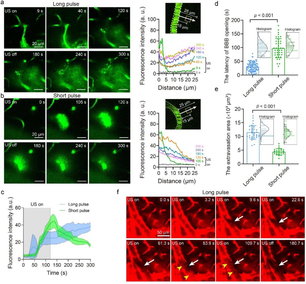
Figure 5. Two-photon image sequences of BBB opening using 120 s long- and short-pulse ultrasound. Time-elapsed fluorescence images showing the cerebrovasculature before, during, and after sonication at long-pulse (A) and short-pulse (B) modes. The image acquisition lasted 300 s. Fluorescence intensity along a path 25 μm from the outer wall of the vessel (indicated by the arrows) were measured under both modes. (C) Quantification of dextran-diffusion fluorescence intensity changes with time from the rectangular regions measuring 15 μm perpendicular to the vessel walls in panel a and b. The mean values are indicated by the solid lines while the shadow regions depict the standard deviation. (D) The latency of BBB opening. (E) The calculated average dextran extravasation area from a single vessel from each image field. (F) In vivo two-photon imaging of endothelial cell loss during long-pulse ultrasound-induced BBB opening in Tek-iCre: Ai 14 mice. A significant loss of tdTomato fluorescence from endothelial cells (white arrows) at 9.6 s from ultrasound application occurred, with no recovery detected at 180.7 s. Transient flow of sediment-like debris (yellow arrowheads) within the vasculature is observed at 83.9 s, subsequently disappearing.
深圳湾实验室分子生理学研究所李芬芳研究员为论文最后通讯作者,广东工业大学马琳教授为本文的共同通讯作者,深圳大学沈圆圆教授、课题组硕士生乔超峰及蔡翼聪为共同第一作者。该研究得到了国家自然科学基金、广东省珠江人才计划、广东省自然科学基金及深圳湾实验室启动经费等项目的支持。
李芬芳课题组长期招收以下方向博士后和博士生:1)声学微流控,空化流体力学;2)治疗超声、超声成像; 3)生物物理。欢迎联系课题组PI: fenfang.li@szbl.ac.cn。课题组网站信息详见https://liff-lab.szbl.ac.cn/research/。
论文标题:
Cross-scale mechanistic insights into ultrasound pulse-length dependent BBB opening
供稿 | 李芬芳课题组
编辑 | 鲍 啦
责编 | 远 山
欢迎投稿、建议 | media@szbl.ac.cn




 粤公网安备44031102000926号